How to Correct Bow Legs in Adults? Treatment Options, Exercises & Surgery
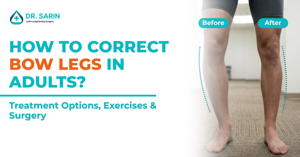
Many adults live with bow legs for years without realizing that treatment options are available. Some have had the condition since childhood. Others develop it gradually due to arthritis, bone disorders, or previous injuries. At first, the issue may seem purely cosmetic. However, as time passes, many people begin noticing symptoms such as knee pain, difficulty walking, uneven posture, and reduced mobility. This naturally leads to an important question: How to correct bow legs in adults? The answer depends on the severity of the deformity, its underlying cause, and the overall health of the knee joint. In this guide, we’ll explain what can and cannot correct bow legs in adults and when professional treatment may be necessary. Can Bow Legs Be Corrected in Adults? Yes. Bow legs can be corrected in adults. However, it’s important to understand that adult bones have finished growing. Unlike children, adults cannot rely on natural growth to straighten the legs. The treatment approach therefore focuses on improving alignment, reducing symptoms, and correcting the underlying deformity. The best solution depends on: The severity of bowing Presence of arthritis Bone structure Age Activity level Overall joint health What Causes Bow Legs in Adults? Before discussing treatment, it’s important to identify the cause. Adult bow legs may develop due to: Untreated childhood bow legs Osteoarthritis Blount’s disease Vitamin D deficiency Rickets Previous fractures Bone deformities Genetic conditions Understanding the root cause helps determine the most effective treatment plan. Can Exercise Correct Bow Legs? This is one of the most common questions patients ask. The honest answer is: Exercise cannot straighten bones that are already misaligned. Many websites claim that stretching or strengthening exercises can completely correct bow legs. Unfortunately, this is not true for adults. Once bone growth is complete, exercises cannot reshape the bones. However, exercise can still provide important benefits. It may help: Improve muscle strength Increase flexibility Support knee stability Reduce discomfort Improve walking mechanics Think of exercise as a supportive treatment rather than a permanent correction. Exercises That May Help Manage Symptoms Although they won’t straighten the legs, certain exercises may improve overall function. These include: Quadriceps Strengthening: Strong thigh muscles provide better support to the knee joint. Hamstring Stretching: Improved flexibility can reduce strain on surrounding structures. Hip Strengthening Exercises: Strong hip muscles help improve overall lower limb mechanics. Low-Impact Activities: Activities such as: Swimming Cycling Walking on flat surfaces can help maintain mobility while reducing stress on the knees. Always consult a specialist before starting any exercise program if significant deformity exists. Can Braces Correct Bow Legs in Adults? Braces are sometimes recommended to improve comfort and stability. However, adults should understand an important limitation: Braces generally do not permanently correct bow leg deformities after skeletal maturity. They may provide: Temporary support Improved walking comfort Better knee alignment during activity But they cannot change the shape of the bones. When Is Treatment Necessary? Not everyone with bow legs requires treatment. However, medical evaluation is recommended if you experience: Knee pain Progressive deformity Walking difficulties Joint stiffness Reduced physical activity Uneven leg alignment Early signs of arthritis The earlier the condition is assessed, the easier it may be to prevent further joint damage. How Are Bow Legs Evaluated? An orthopedic specialist typically performs: Physical Examination This helps assess: Leg alignment Walking pattern Knee stability Joint movement Standing X-Rays These images reveal: Severity of bowing Joint wear Bone structure Alignment abnormalities Accurate imaging is essential before deciding on treatment. Non-Surgical Treatment Options Mild bow leg deformities may be managed conservatively. Weight Management: Excess body weight increases pressure on already stressed knee joints. Maintaining a healthy weight may help reduce symptoms. Physical Therapy: A structured rehabilitation program may improve: Strength, Flexibility, Balance, and Walking mechanics. Pain Management: Depending on symptoms, treatment may include: Anti-inflammatory medications, Lifestyle modifications, and Activity adjustments. While these approaches help manage discomfort, they do not correct the underlying bone alignment. When Is Surgery Recommended? Surgery may be considered when: Bowing is severe Knee pain becomes significant Walking is affected Arthritis is developing Conservative treatments fail The goal is not simply cosmetic improvement. The primary objective is restoring proper alignment and protecting the knee joint from further damage. Surgical Correction for Bow Legs Modern orthopedic correction procedures can effectively straighten the legs and restore mechanical alignment. Bow legs correction surgery may help: Improve leg alignment Reduce knee stress Relieve pain Improve posture Enhance mobility Slow joint degeneration For many adults with moderate to severe deformities, surgery offers the most predictable long-term solution. What Is Recovery Like After Bow Leg Correction? Recovery varies depending on: Type of procedure Severity of deformity Patient health Rehabilitation progress Most treatment plans include: Guided physiotherapy Progressive weight-bearing Strengthening exercises Regular follow-up evaluations The goal is to safely restore function while achieving optimal alignment. What Happens If Bow Legs Are Left Untreated? Ignoring significant bow legs can lead to ongoing stress on the knee joint. Potential complications include: Progressive arthritis Chronic knee pain Joint degeneration Reduced mobility Difficulty walking Poor posture Early intervention often helps prevent these long-term issues. Can Adults Achieve Normal-Looking Legs After Treatment? In many cases, yes. Modern orthopedic correction techniques are designed to improve both function and appearance. Many patients experience: Straighter leg alignment Better walking mechanics Reduced discomfort Improved confidence Enhanced quality of life The exact outcome depends on the severity of the deformity and the chosen treatment approach. Final Thoughts: How to Correct Bow Legs in Adults? If you’re wondering how to correct bow legs in adults, the most important step is identifying the underlying cause and severity of the condition. While exercises and therapy can help manage symptoms, they cannot permanently straighten adult bones. For individuals with significant deformity, persistent pain, or worsening alignment, professional orthopedic evaluation is essential. With modern treatment options, many adults can achieve improved alignment, better mobility, and long-term joint health. The sooner the condition is assessed, the better the opportunity to protect your knees and maintain an active lifestyle. FAQs About Bow Legs Correcton in Adults?
What Causes Bow Legs in Adults? Understanding the Real Reasons Behind Leg Misalignment
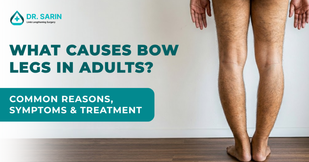
Many people believe bow legs are only a childhood condition. However, that’s not always true. Some adults continue to have bow legs from childhood, while others develop the condition later in life due to joint problems, injuries, or bone disorders. If you’ve noticed your knees staying apart when your feet are together, or you’ve started experiencing knee pain along with a curved leg appearance, you may be wondering: What causes bow legs in adults? Understanding the underlying cause is important because bow legs can affect much more than appearance. Left untreated, the condition may lead to pain, mobility issues, and even early arthritis. In this guide, we’ll explore the common causes of bow legs in adults, associated symptoms, and available treatment options. What Are Bow Legs? Bow legs, medically known as Genu Varum, occur when the legs curve outward, creating a noticeable gap between the knees while the ankles remain close together. This abnormal alignment changes how body weight is distributed through the knee joints. Instead of being evenly balanced, excessive pressure is placed on the inner side of the knees. Over time, this imbalance can affect walking, posture, and joint health. Know More: What is Bow Legs? Can Adults Develop Bow Legs? Yes, While some adults have had bow legs since childhood, others develop the condition later in life. Adult bow legs often result from: Joint degeneration Previous injuries Bone diseases Growth abnormalities Arthritis-related changes In many cases, the deformity gradually worsens if the underlying cause remains untreated. What Causes Bow Legs in Adults? Several medical conditions can contribute to bow leg deformity in adulthood. Let’s look at the most common causes. 1. Untreated Childhood Bow Legs One of the most common causes is a bow leg condition that never fully corrected during childhood. Some individuals had: Physiologic bowing Blount’s disease Rickets Growth plate abnormalities Although symptoms may have been mild earlier in life, the deformity often becomes more noticeable with age. As body weight increases and joints experience more wear and tear, knee pain and mobility issues can develop. 2. Knee Osteoarthritis Osteoarthritis is among the leading causes of bow legs in adults. Over time, the cartilage inside the knee gradually wears away. When the inner portion of the knee deteriorates more rapidly than the outer side, the leg begins shifting outward. This creates a bow-legged appearance. Common signs include: Knee pain Stiffness Swelling Difficulty walking Reduced mobility Many adults seeking treatment for bow legs are actually experiencing advanced osteoarthritis-related deformity. 3. Blount’s Disease Blount’s disease affects the growth plate near the upper part of the shin bone (tibia). Although commonly diagnosed in childhood, untreated cases can persist into adulthood. As growth continues abnormally, the lower legs gradually curve outward. Adults with untreated Blount’s disease often experience: Progressive bowing Knee pain Walking difficulties Joint instability 4. Rickets and Vitamin D Deficiency Healthy bones require adequate amounts of: Vitamin D Calcium Phosphorus When these nutrients are deficient, bones can become soft and weak. In severe cases, the bones may bend under the body’s weight, leading to bow leg deformities. Although rickets is more common in children, adults with long-standing bone weakness can continue experiencing alignment problems. 5. Bone Fractures That Heal Improperly A previous fracture involving the thigh bone (femur) or shin bone (tibia) may cause bow legs if the bone heals in an incorrect position. This is known as a malunion. Even a small alignment error can alter weight distribution across the knee joint. Over time, this may lead to: Visible bowing Uneven walking Joint pain Increased risk of arthritis 6. Paget’s Disease of Bone Paget’s disease is a chronic bone disorder that affects the normal process of bone remodeling. The bones become enlarged, weaker, and structurally abnormal. When the disease affects the leg bones, bowing may occur. Symptoms often include: Bone pain Joint discomfort Leg deformity Reduced mobility Although less common, Paget’s disease remains an important cause of bow legs in older adults. 7. Genetic Bone Disorders Certain inherited conditions can affect bone growth and alignment. These disorders may alter: Bone shape Growth patterns Joint structure As a result, bow leg deformities can persist into adulthood. A detailed orthopedic evaluation is usually required to identify these underlying conditions. How Do Bow Legs Affect Adults? Many adults initially view bow legs as a cosmetic issue. However, the condition can have significant functional consequences. Because the knees are not properly aligned, excessive pressure develops inside the joint. This can lead to: Chronic knee pain Walking difficulties Reduced stamina Joint degeneration Postural imbalance Hip discomfort Lower back pain The longer the misalignment persists, the greater the stress placed on surrounding joints. Symptoms That Should Not Be Ignored You should consider medical evaluation if you experience: Progressive leg bowing Knee pain while walking Difficulty climbing stairs Joint stiffness Uneven walking pattern Reduced physical activity due to discomfort Early diagnosis often helps prevent further joint damage. How Are Bow Legs Diagnosed in Adults? Diagnosis typically involves a combination of: Physical Examination An orthopedic specialist evaluates: Leg alignment Walking pattern Joint movement Overall posture X-Rays Standing X-rays help determine: Severity of deformity Knee alignment Joint damage Bone abnormalities These images are essential for treatment planning. Can Bow Legs in Adults Be Corrected? Yes, Modern orthopedic techniques can effectively correct bow leg deformities in adults. The most appropriate treatment depends on: Cause of the deformity Severity of misalignment Age Joint health Presence of arthritis Treatment Options for Adult Bow Legs Non-Surgical Treatment Mild cases may benefit from: Physical therapy Weight management Activity modification Pain management strategies However, these treatments generally do not correct the actual bone alignment. Surgical Correction When significant deformity exists, Bow legs correction surgery may be recommended. The goals of surgery include: Restoring proper leg alignment Improving walking mechanics Reducing knee stress Preventing further joint damage Improving quality of life Advanced corrective procedures can provide long-term functional and cosmetic improvements. What Happens If Adult Bow Legs Are Left Untreated? Without proper treatment, severe bow leg deformities may continue worsening. Potential
What Is Bow Legged? Understanding the Condition, Causes, and Treatment Options
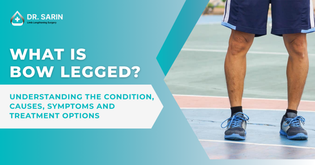
Have you ever noticed someone’s legs curve outward while standing with their feet together? This condition is commonly known as bow legged, medically referred to as Genu Varum. Many people assume bow legs are only seen in babies and young children. While this is often true, bow legs can also persist into adulthood or develop later in life due to underlying medical conditions. The good news is that modern orthopedic treatments can successfully correct bow leg deformities and improve both appearance and function. In this guide, we’ll explain what bow legged means, why it happens, and when treatment may be necessary. What Does Bow Legged Mean? A person is considered bow legged when the legs curve outward at the knees while the ankles remain close together. When standing with both feet together: The ankles touch The knees remain apart The legs create a noticeable bow-like shape This condition is known medically as Genu Varum. In mild cases, the curvature may be barely noticeable. In more severe cases, it can affect walking, posture, balance, and knee function. Is Bow Legged Normal? The answer depends on age. In infants and toddlers, bow legs are often a normal part of development. Most babies are born with a slight outward curvature of the legs because of their position inside the womb. As children grow and begin walking, the legs gradually straighten. For many children: Bow legs improve naturally by age 2-3 years No treatment is required Regular observation is usually sufficient However, if the condition persists beyond early childhood or becomes progressively worse, further evaluation may be necessary. What Causes Bow Legs? Several factors can lead to bow leg deformity. Natural Growth and Development: This is the most common reason in young children. As the child’s bones develop, the legs often straighten naturally without intervention. Rickets: Rickets occurs when bones become soft due to a deficiency of Vitamin D, calcium, or phosphate. This can cause: Weak bones, Growth abnormalities, and Bowing of the legs. Blount’s Disease: Blount’s disease affects the growth plate near the knee and can cause progressive bowing. Without treatment, the deformity may worsen over time. Bone Injuries: Fractures that heal improperly can lead to uneven bone alignment and bow leg formation. Arthritis. In adults, knee arthritis can gradually change joint alignment and result in bow-legged appearance. Genetic Conditions: Certain inherited bone disorders can affect bone growth and alignment, leading to genu varum. Common Symptoms of Bow Legs Some individuals experience only cosmetic concerns, while others develop functional problems. Common symptoms include: Outward curvature of the legs Knees that remain apart when standing Altered walking pattern Knee pain Hip discomfort Reduced balance Difficulty walking long distances As the condition progresses, abnormal pressure on the knee joint can increase the risk of arthritis. How Is Bow Legged Diagnosed? Diagnosis usually begins with a physical examination. An orthopedic specialist may evaluate: Leg alignment Walking pattern Knee positioning Joint movement Imaging studies such as X-rays help determine: Severity of the deformity Bone alignment Underlying causes Accurate diagnosis is essential because treatment depends on the root cause. When Should Bow Legs Be Treated? Treatment is not always necessary. Medical evaluation is recommended when: Bow legs worsen over time The child is older than 3 years and the condition persists One leg is more curved than the other Pain develops Walking becomes difficult The deformity affects daily activities Early intervention can help prevent future complications. Treatment Options for Bow Legs Bow Legs Treatment varies depending on age, severity, and underlying cause. Observation: Many young children simply require monitoring because the condition often improves naturally. Nutritional Management: If Vitamin D deficiency or rickets is responsible, correcting the deficiency may help improve bone health. Bracing: In selected pediatric cases, braces may help guide proper growth and alignment. Surgical Correction: Adults and patients with significant deformities often require surgical correction. Modern procedures can: Straighten the legs, Improve walking mechanics, Reduce knee stress, Improve posture, Enhance confidence Advanced corrective procedures are designed to restore proper alignment while preserving long-term knee function. What Happens If Bow Legs Are Left Untreated? Severe bow leg deformities may increase stress on the inner side of the knee joint. Over time, this can contribute to: Early arthritis Chronic knee pain Joint degeneration Walking difficulties Reduced mobility This is why persistent or worsening bow legs should not be ignored. Can Adults Correct Bow Legs? Yes. Contrary to popular belief, bow legs can be corrected in adults. Since adult bones have completed growth, treatment usually focuses on orthopedic correction procedures that realign the bones and restore proper leg mechanics. Modern techniques provide highly predictable outcomes for suitable candidates. Final Thoughts Understanding what is bow legged is the first step toward recognizing whether the condition requires treatment. While mild bowing in young children is often normal, persistent or severe bow legs can affect mobility, posture, knee health, and overall quality of life. If bow legs continue beyond childhood or begin causing discomfort, a consultation with an experienced orthopedic specialist can help determine the most appropriate treatment approach. Early evaluation and proper management can significantly improve both function and long-term joint health. Bow Legged FAQs 1. What is bow legged? Ans: Bow legged, or Genu Varum, is a condition where the legs curve outward, causing the knees to remain apart when the ankles are together. 2. Are bow legs normal in children? Ans: Yes. Mild bow legs are common in infants and toddlers and often correct naturally as the child grows. 3. What causes bow legs in adults? Ans: Common causes include arthritis, previous bone injuries, untreated childhood deformities, and certain bone disorders. 4. Can bow legs be corrected without surgery? Ans: Some childhood cases improve naturally or respond to bracing and nutritional treatment. Adults with significant deformities often require surgical correction. 5. Do bow legs cause knee pain? Ans: Yes. Severe bow legs can place extra stress on the knee joint, leading to pain, instability, and early arthritis over time.
Precice vs Ilizarov vs LON Method Comparison Guide
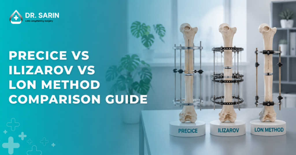
Choosing the right technique is one of the most important decisions in limb lengthening surgery. Many patients feel confused when they hear about different options like Precice, Ilizarov, and LON method. If you are planning height increase surgery, understanding the differences between these methods will help you make a better and safer choice. Each technique has its own advantages, limitations, and suitability depending on the patient’s condition. In this guide, we will explain all three methods in a simple way so you can clearly understand which one may be best for you. Overview of Limb Lengthening Methods All methods of limb lengthening surgery work on the same basic principle. The bone is carefully cut and then gradually lengthened so that new bone forms in the gap. However, the difference lies in: how the bone is lengthened what type of device is used comfort level during recovery You may have already explored some basics in Available Methods Of Limb Lengthening, but here we will go deeper into comparison. What Is the Ilizarov Method? The Ilizarov method is one of the oldest and most widely used techniques. How it works?: External metal rings are fixed around the leg Pins and wires are attached to the bone The device is adjusted daily to increase length Advantages: suitable for complex cases strong and reliable method widely used for medical conditions Disadvantages: visible external frame higher discomfort compared to internal methods risk of pin site infection Despite being older, it is still considered very effective. You may also relate this with Why Ilizarov Technique Is Still the Gold Standard in Complex Cases? What Is the Precice Method? The Precice method is a modern and advanced technique used in height increase surgery. How it works: a magnetic rod is inserted inside the bone lengthening is controlled using an external remote no external frames are used Advantages: no visible external device more comfortable for patients lower risk of infection better cosmetic appearance Disadvantages: higher cost not suitable for all cases requires advanced medical setup This method is commonly preferred in limb lengthening surgery in Delhi for patients looking for comfort and better aesthetics. What Is the LON Method (Lengthening Over Nail)? The LON method is a combination of both internal and external techniques. How it works: an internal rod is inserted into the bone an external fixator is used temporarily after lengthening, the external device is removed Advantages: shorter time with external device faster recovery compared to Ilizarov more stable than fully external methods Disadvantages: still involves external device for some time moderate risk of infection more complex procedure This method is often considered a balanced option between comfort and cost. Precice vs Ilizarov vs LON: Key Differences 1. Comfort Level Precice: highest comfort LON: moderate comfort Ilizarov: lowest comfort 2. Visibility Precice: no external device LON: temporary external device Ilizarov: fully external frame 3. Cost Precice: most expensive LON: mid-range Ilizarov: more affordable You can also explore Factors That Can Influence the Price of Limb Lengthening Surgery in India for deeper insights. 4. Recovery Experience Precice: smoother recovery LON: moderate recovery Ilizarov: longer and more challenging This connects with topics like Limb Lengthening Surgery Recovery Timeline Guide. 5. Risk of Infection Precice: lowest LON: moderate Ilizarov: higher due to external pins Which Method Is Best for You There is no single “best” method for everyone. Precice is suitable if: you want better comfort you prefer no visible device budget is not a major issue Ilizarov is suitable if: you have a complex medical condition cost is a concern strong correction is needed LON is suitable if: you want a balance of cost and comfort you are okay with temporary external support Choosing the right method depends on your body condition and goals. This is why consultation before height increase surgery in Delhi is very important. Role of Surgeon in Method Selection The success of limb lengthening surgery depends not just on the method, but also on the surgeon’s expertise. An experienced surgeon will: evaluate your bone condition suggest the safest method guide you through recovery This is especially important in advanced centers offering limb lengthening surgery in Delhi. Things to Consider Before Choosing Before selecting a method, think about: your comfort preference budget recovery time medical condition You should also read Selecting the Ideal Limb Lengthening Approach: Comparing Femur and Tibia Lengthening to understand more factors. Long-Term Results of Each Method All three methods can provide good results if done correctly. Patients usually achieve: increased height improved body balance better confidence You may also connect this with Long-Term Effects Of Limb Lengthening Surgery. Final Thoughts Each method-Precice, Ilizarov, and LON-has its own place in limb lengthening surgery. The right choice depends on your personal needs, medical condition, and expectations. Modern advancements in height increase surgery have made all these options safer and more effective than before.
How to Lengthen Legs Naturally & Surgically in 2026: A Complete Guide to Getting Taller
Want to Know How to Lengthen Legs? Let’s be honest, many of us have looked in the mirror and wished we had longer legs. Whether it’s for confidence, better body proportions, or just to feel taller in everyday life, the desire is real. The good news? You’re not stuck with what you’ve got. From expert surgical options to lifestyle adjustments, this guide walks you through how to lengthen legs effectively and safely, whether you’re 18 or 38. And if you’re serious about results, there’s no one better than Dr. Amar Sarin, Asia’s leading limb lengthening surgeon. How to Get Longer Legs: The Top 3 Paths 1. Limb Lengthening Surgery (The Permanent, Scientific Way) If you’ve tried everything and still haven’t seen changes, Limb Lengthening Surgery is the most effective and lasting solution. Under the guidance of Dr. Amar Sarin, this advanced medical procedure carefully stretches the leg bones, helping you gain 2–6 inches in height. Why choose Dr. Amar Sarin? 30+ years of experience 3000+ surgeries performed Recognized as the most experienced and successful Limb Lengthening Surgeon in Asia The procedure uses cutting-edge devices and methods like the Ilizarov technique. The process is gradual and safe, with full recovery monitored by a dedicated team. Whether you’re addressing a medical need or simply want to grow taller for confidence, this is the gold standard solution. For more detailed read “Why Choose Us?“ Book an appointment with Dr. Amar Sarin, Now! 2. Targeted Exercises to Improve Posture & Leg Appearance While exercise won’t make your bones longer, it can improve your posture and muscle tone, making your legs look longer. Here are some go-to moves: Hanging stretches: decompress your spine and hips Leg raises: strengthen your quads and make legs appear leaner Pilates or Yoga: improves posture and alignment Swimming: elongates the body over time Consistent stretching can help optimize your current height, especially if you’ve got bad posture. 3. Fashion Tricks to “Fake” Longer Legs While it’s not a real fix, let’s be real, we all love quick wins. Try these: Wear high-waisted pants Match shoes to your skin tone (or pant color) Go for vertical patterns Avoid cropped pants or ankle straps You’ll be surprised how much taller you can look with these subtle style hacks. How to Increase Length (for Real) Wondering how to increase the length of your legs in a way that actually works? Let’s break it down into what’s temporary vs. what’s permanent: Method Permanent? Average Gain Time to Results Limb Lengthening Surgery ✅ Yes 2–6 inches 6–12 months Exercise & Stretching ❌ No None (visual) Ongoing Fashion Tricks ❌ No Illusion only Instant If you’re serious about how to lengthen legs, surgery with a trusted expert like Dr. Amar Sarin is your best bet. Why Dr. Amar Sarin Is the Best Choice When choosing a surgeon, experience matters. Here’s why Dr. Sarin stands out: Asia’s Most Experienced Limb Lengthening Surgeon Over 3000+ successful surgeries using the Ilizarov method Expert in both cosmetic and corrective limb lengthening Known for treating leg length discrepancies, dwarfism, polio deformities, and more Trusted by international patients from across the world He operates at Sri Balaji Action Medical Institute, equipped with state-of-the-art facilities and a compassionate team dedicated to your recovery. How to Get Started: Your Next Steps Here’s how you can take the first step toward longer legs: Book a Consultation: Speak with Dr. Amar Sarin to understand your goals Undergo Evaluation: Get imaging and physical assessments Discuss Your Options: Understand surgical plans, timelines, and recovery Begin Your Journey: With expert guidance, start your limb lengthening procedure Book your consultation now! Conclusion: Yes, You Can Lengthen Your Legs It’s no longer a dream. Whether you’re aiming to correct a medical issue or simply want to increase your height for confidence, there’s a safe, proven answer to how to lengthen legs, and it starts with expert care. With Dr. Amar Sarin’s unmatched experience and the life-changing possibilities of Height Increase lengthening surgery, you don’t just stand taller, you walk into a new life. Your journey to new heights begins now. Frequently Asked Questions Q: Is it really possible to lengthen legs after 18? Yes. Limb Lengthening Surgery can increase leg length in adults well beyond puberty. Q: How long does recovery take? Complete recovery may take 6–12 months, including physical therapy and bone healing. Q: Is the surgery painful? Mild discomfort is expected, but pain is well-managed with proper care and medications. Q: Can exercises make your legs longer? Not permanently. But they can improve posture and visual length. Q: How can I get started with the surgery? Book a consultation with Dr. Amar Sarin to get personalized advice and a treatment plan. Related Blogs: Limb Lengthening Surgery in 2026: Latest Technologies, Costs, and Recovery Insights Limb Lengthening Surgery in India | Why International Patients Choose Dr. Sarin What Conditions Can Limb Lengthening Surgery Effectively Treat? Can I Still Run After Height Increase Surgery? Why Height Increases Stop After 18?
Limb Lengthening Surgery in 2026: Latest Technologies, Costs, and Recovery Insights
In 2026, limb lengthening surgery is more advanced, safer, and in higher demand than ever. With innovations in medical technology, improved recovery timelines, and growing patient success stories, individuals worldwide are turning to cosmetic height surgery to transform their lives. At the forefront of this revolution is Dr. Amar Sarin, Asia’s leading authority in limb lengthening, with 30+ years of experience and over 3,000 successful procedures. Why Are More People Opting for Limb Lengthening Surgery in 2026? Cosmetic height surgery is no longer taboo patients seek it to: Gain confidence in professional and personal life Improve body proportions Correct physical deformities or post-polio conditions With advancements in minimally invasive techniques and AI-controlled devices, the process has become more predictable, accessible, and life-changing. What’s New in Limb Lengthening Surgery in 2026? 1. Advanced Internal Lengthening Devices Modern systems like PRECICE 2.2 offer: Remote-controlled lengthening with magnetic fields No need for external fixators Minimal pain and lower infection risk Dr. Sarin also uses hybrid techniques like LON (Lengthening Over Nail) for added safety and stability during tibial or femoral lengthening. 2. Scar-Minimizing Surgical Protocols Small incisions and bone-preserving techniques help reduce scarring and speed up healing. 3. Smart Monitoring Systems Limb lengthening procedures at Dr. Sarin’s center are supported by advanced smart monitoring systems used exclusively by the medical team. These allow for: Real-time tracking of bone distraction progress Immediate adjustment of distraction rates if needed Timely detection of complications like premature consolidation or delayed union Patients benefit from close, in-person supervision during regular checkups, ensuring precision and safety throughout the lengthening phase. Limb Lengthening Surgery Cost: India vs. Global (2026) Region Average Cost (Both Legs) What’s Included India (Dr. Sarin) ₹16–30 lakhs ($18K–35K) Surgery, hospital stay, rehab, support USA $85,000–$155,000 Surgery only (excludes rehab) Germany/S. Korea $50,000–$90,000 Advanced rehab plans included Note: India is the most cost-effective location globally for advanced limb lengthening procedures. Week-by-Week Recovery Timeline Week 1–2: Post-Surgery Stay Hospitalization at Sri Balaji Action Medical Institute Light walking with walker Daily monitoring by Dr. Sarin’s team Week 3–8: Lengthening Phase 1 mm/day bone distraction Adjusted with mobile app Physiotherapy under Shariq Mohammed, rehab expert Month 3–6: Bone Hardening Return to normal walking Upper-body gym begins Driving possible 6–12 Months: Full Function Return Sports resumption Return to travel, light jogging 12–24 Months: Nail Removal Final step to complete recovery Read more: Timeline for Lengthening and Recovery Common Patient Questions Q. Is limb lengthening painful? Ans: The lengthening itself is painless, though some muscle tightness and discomfort are expected. Pain is managed with medication and physiotherapy. Q. How safe is the surgery now? Ans: With zero fat embolism cases at Dr. Sarin’s practice and 3,000+ successful operations, the procedure is extremely safe when performed by experienced professionals. Q. Can I run or play sports again? Ans: Yes. Patients usually resume athletic activities within 12 months and regain full sports capability by 24 months. Q. Why Choose Dr. Amar Sarin? 30+ years of expertise Performed 3,000+ Ilizarov and internal lengthening surgeries Recognized by major outlets: Times of India, Hindustan Times, CBC Full pre-surgery, post-op, and in-house rehab care Supported by a top team including: Dr. Sunil Kumar, FRCS (Glasgow), MCh (Liverpool) Dr. Yogesh Kumar, Orthopaedic Surgeon & DNB trainer Dr. Govind Vallabh Joshi, DNB Ortho, published researcher See Real Results Check our before-and-after gallery: View Results » Success stories from India, Iraq, the UAE, and beyond. Booking & Contact Details Sri Balaji Action Medical Institute, New Delhi Rehab Centre – DLF Phase 1, Gurugram Contact Number: +91 9810376803 amarsarin@hotmail.com Book Your Consultation Now » You May Also Like to Read: Average Height of UK 2026 Best Diet for Bone Healing After Limb Lengthening How We Help You Return to Work and Sports After Limb Lengthening? How to Lengthen Legs Naturally & Surgically in 2026 Limb Lengthening Surgery in India
Average Height of UK 2026

The average height of a population serves as a key indicator of overall health and nutrition levels within a country. In the UK, this metric has shown a steady increase over the years, driven by improvements in living conditions, advancements in healthcare, and enhanced dietary habits. Understanding these trends not only provides insight into public health but also informs policies aimed at fostering a healthier future. Current Average Height of UK (2026) As of 2025, the average height for adults in the UK is as follows: These figures represent a gradual rise over recent decades, reflecting the positive impact of various societal factors. Factors Affecting Average Height in the UK Several elements contribute to the average height of individuals in the UK: Historical Trends in Height Over the past century, historical data indicates a consistent increase in average height across the UK. This trend can be attributed to enhanced living standards, medical advancements, and improved nutrition. Notably, the post-World War II era saw significant improvements in public health policies, which have had lasting effects on population height. Period Approx. Average Height (Men) Height in cm Medieval (~1400–1700) Similar to 20th century ~170–172 cm Late 18th century ~5 ft 6 in ~167 cm Mid 19th century 5 ft 7–8 in 170–173 cm 1900 — ~172 cm 1950 — ~175 cm 1970 — ~177 cm Recent (men) ~5ft 9 in ~175 cm Recent (women) ~5ft 4 in ~162 cm Regional Variations While national averages provide a broad overview, there are regional variations in average height within the UK. These differences can be influenced by local dietary habits, socioeconomic conditions, and genetic diversity. For example, studies have shown that individuals in urban areas may have different average heights compared to those in rural regions, often due to lifestyle and dietary differences. The Impact of Global Trends In addition to local factors, global trends such as increased migration and cultural exchange can influence dietary habits and health practices, potentially affecting average height. As the UK becomes more multicultural, the blending of dietary practices may lead to new health outcomes. Conclusion Understanding the average height and the factors that influence it is essential for developing policies aimed at improving public health and nutrition. By addressing these areas, we can continue to support the positive trend in average height and overall well-being in the UK. As we move forward, ongoing research and community engagement will be crucial in ensuring that all individuals have the opportunity to reach their full height potential, contributing to a healthier society overall. Additional Resources For those interested in exploring this topic further, consider the following resources: NHS Guidelines on Nutrition: NHS Nutrition Public Health England Reports on Height Trends: PHE Reports Research on Genetics and Height: Genetics Home Reference You May Also Like to Read: Best Diet for Bone Healing After Limb Lengthening How We Help You Return to Work and Sports After Limb Lengthening? How to Lengthen Legs Naturally & Surgically in 2026 Limb Lengthening Surgery in 2026 Limb Lengthening Surgery in India
How Safe Is Limb Lengthening Surgery? Risks and Precautions

Many people who want to increase their height often ask one simple question: “Is limb lengthening surgery safe?” As a surgeon who performs height increase surgery regularly, I understand why patients are concerned. Any procedure that changes bone structure naturally raises questions about safety, risks, side effects, and cost. Here I’m trying to clear all your doubts about this surgery, I will explain how safe limb lengthening surgery is, the possible risks, and what patients should know before considering height increasing surgery. What Is Limb Lengthening Surgery? Limb lengthening surgery is a medical procedure used to increase a person’s height by gradually lengthening the bones in the legs. Many people also refer to it as: Height increase surgery Bone lengthening surgery Leg lengthening surgery Many patients often ask questions like: Can I increase height by surgery? Is there any surgery to increase height? What is the height increasing surgery name? and the answer of all these queries is: Yes, the procedure exists and is commonly known as limb lengthening surgery. During the surgery, the bone is carefully cut and gradually lengthened using a specialized medical device. Over time, new bone forms in the gap, allowing the leg to grow longer. Most patients can increase their height by 5-8 cm (2-3 inches) through this process. Is Limb Lengthening Surgery Safe? A very common question I hear from patients is: “Is limb lengthening surgery safe or not?” The answer is that limb lengthening surgery can be safe when performed by an experienced orthopedic surgeon with proper medical supervision. From my clinical experience, most patients recover well when the surgery is done correctly and when they follow post-surgery physiotherapy and precautions. The Possible Limb Lengthening Surgery Risks Some possible risks of limb lengthening surgery include: Infection: Infections can sometimes occur around the surgical area or fixation device. These are usually treatable with medication if detected early. Nerve Stretching: During the lengthening process, nerves may experience stretching, which can cause temporary numbness or tingling. Joint Stiffness: If physiotherapy is not done regularly, the knee or ankle joint may become stiff. Bone Healing Issues: In rare cases, the new bone may take longer to grow or may need additional support. These are the reasons why patients often ask “how risky is limb lengthening surgery?” Proper surgical planning and monitoring help minimize these risks. Limb Lengthening Surgery Side Effects Some of the side effects of limb lengthening surgery may include: Pain during the lengthening phase Muscle tightness in the legs Temporary swelling Difficulty walking during early recovery In rare cases, leg lengthening complications may occur, but careful follow-up and physiotherapy help manage most issues. Can Height Surgery Go Wrong? Patients sometimes ask: “Can height surgery go wrong?” Complications can happen in any surgical procedure, especially if: The surgery is done by an inexperienced surgeon The patient does not follow rehabilitation instructions The bone is lengthened too quickly That is why choosing the right surgeon and hospital is extremely important for safe limb lengthening surgery. Limb Lengthening Surgery Cost in India The limb lengthening surgery cost in India can vary depending on factors like the surgical technique used, hospital facilities, surgeon expertise, and post-surgery care. Many patients search for height surgery cost or leg lengthening surgery cost in India, but the final cost differs for each patient and treatment plan. Is Limb Lengthening Surgery Safe in India? Another question patients often ask is: “Is limb lengthening surgery safe in India?” Yes, safe limb lengthening in India is possible when the procedure is performed in specialized orthopedic centers with experienced surgeons and proper rehabilitation support. Before choosing a surgeon, patients should always: Research the doctor’s experience Check patient reviews and outcomes Understand the complete treatment plan Who Can Consider Height Increase Surgery? Height increasing surgery is usually suitable for: Adults who want cosmetic height increase Patients with leg length differences Individuals with certain bone conditions Final Words When performed correctly by an experienced surgeon and followed by proper rehabilitation, limb lengthening surgery can be a safe and effective way to increase height. However, it is important to understand the risks of limb lengthening surgery, possible side effects, and the recovery process before making a decision. Consulting a qualified specialist “Dr. Amar Sarin” will help you determine whether height increase surgery is the right option for you. You May Also Like to Know: Average Height Across India 2026 by Select City How Much Height Can You Gain Safely with Limb Lengthening? Indian children may no longer outgrow parents What’s The Average Height Per Country? Psychological Impact Of Limb-Lengthening Surgery
Limb Lengthening Surgery Recovery Timeline Guide
Recovery is one of the most important parts of limb lengthening surgery. Many people focus only on the surgery itself, but the real results depend on how well your body heals over time. If you are planning height increase surgery, understanding the full recovery timeline will help you stay prepared and confident throughout the journey. Overview of Recovery Process Recovery after limb lengthening surgery happens in different stages. Each stage has its own purpose, challenges, and progress. The total recovery time can take several months, depending on: The amount of height gained Your bone healing speed Overall health Advanced care during limb lengthening surgery in Delhi helps make recovery more structured and safer. Stage 1: First 1–7 Days (Initial Recovery) This is the immediate phase after surgery. What happens: You stay under medical supervision Pain and swelling are managed Movement starts slowly Doctors focus on stability and preventing complications. This phase is similar to what is explained in Guide to the Initial 10 Days Following Leg Lengthening Surgery. Stage 2: Week 1 to Week 6 (Lengthening Phase) This is the most important stage. What happens: The bone is slowly lengthened Small adjustments are made daily New bone starts forming Patients may feel tightness or discomfort, which is normal. During this time: Physiotherapy is very important Regular follow-ups are required You can also relate this stage with Discover the Phases of Limb Lengthening for better understanding. Stage 3: Month 2 to Month 4 (Early Bone Healing) In this stage, the new bone becomes stronger. What happens: Bone gap starts filling Mobility slowly improves Pain reduces This phase is strongly connected with Bone Healing After Leg Lengthening Surgery. Nutrition also plays a key role here. Proper diet supports faster healing, as explained in Bone Recovery Post Limb Lengthening Surgery with Nutrition and Supplements. Stage 4: Month 4 to Month 8 (Consolidation Phase) This is the strengthening stage. What happens: new bone becomes solid walking improves support devices may still be used Patients gradually return to normal daily activities. You can also explore Returning to Work and Daily Life After Limb Lengthening Surgery for more clarity. Stage 5: Month 8 to 12 (Full Recovery Phase) This is the final stage of recovery. What happens: Bones fully heal Normal walking resumes Strength improves Some patients may even return to sports, depending on their condition. You may find this useful along with Can You Do Sports after Limb Lengthening Surgery? Factors That Affect Recovery Time Not everyone heals at the same speed. Recovery after height increase surgery depends on: 1. Age: Younger patients usually heal faster. 2. Bone Health: Strong bones support better regeneration. 3. Nutrition: A healthy diet improves healing speed. 4. Physical Activity: Regular physiotherapy is very important. 5. Surgeon Experience: Choosing experts in limb lengthening surgery in Delhi ensures better care and guidance. Common Challenges During Recovery Recovery is not always easy. Some common issues include: Stiffness in joints Muscle tightness Slow movement initially These are normal and improve with time and proper care. You can also read Dos and Don’ts Of Exercise and Physical Activity Following Limb Lengthening Surgery to avoid mistakes. Tips for Faster Recovery To improve your recovery experience: Follow doctor’s advice strictly Attend all physiotherapy sessions Maintain a healthy diet Avoid stress and overexertion Consistency is the key to success. When Can You Walk Normally Most patients can: Start assisted walking within weeks Walk more independently after a few months Return to normal walking by the end of recovery However, results vary based on individual healing. Final Thoughts The limb lengthening surgery recovery timeline requires patience and commitment. While the process may feel long, each stage brings you closer to your final result. With proper care, guidance, and modern treatment options in limb lengthening surgery in Delhi, recovery can be smooth and successful. Recovery is not just about healing – it is about rebuilding strength and confidence. If you are planning to increase height, knowing what to expect can make your journey easier. Stay consistent, follow expert advice, and trust the process.
Is Limb Lengthening Surgery Worth It?
Many people who are unhappy with their height often ask one important question: is limb lengthening surgery worth it? With growing awareness about height increase surgery, more individuals are exploring this option. But before making a decision, it is important to understand the benefits, risks, cost, and long-term results. This article will help you make a clear and practical decision. What Is Limb Lengthening Surgery Limb lengthening surgery is a medical procedure that helps increase a person’s height by gradually lengthening the bones of the legs. It works through a process called distraction, where the bone is slowly separated so new bone forms in the gap. Today, advanced techniques used in limb lengthening surgery in Delhi make the procedure safer and more controlled than before. Why Do People Consider This Surgery? There are two main reasons why people choose this surgery: 1. Cosmetic Reasons Some people feel less confident due to short height. For them, height increase surgery can improve self-image and confidence. 2. Medical Conditions In some cases, people have uneven leg length or conditions like dwarfism. Surgery helps correct these problems and improve daily life. Benefits of Limb Lengthening Surgery 1. Increase in Height The most obvious benefit is gaining height. On average, patients can gain a few inches depending on the procedure. 2. Better Confidence Many patients report improved confidence after surgery. This can impact both personal and professional life. You may have already read about this in related topics like Life After Gaining Height: Confidence, Career & Social Impact. 3. Improved Body Balance In medical cases, it helps correct limb differences and improves walking and posture. Risks and Challenges While the benefits are clear, it is also important to understand the risks. 1. Pain and Discomfort During the lengthening phase, patients may feel discomfort. However, proper care and guidance can help manage this. You can also explore Pain Management After Limb Lengthening Surgery for more details. 2. Long Recovery Time Recovery is not quick. It may take several months before normal activities can resume. Topics like Returning to Work and Daily Life After Limb Lengthening Surgery can give you better clarity. 3. Possible Complications Like any surgery, there are risks such as infection or slow bone healing. Choosing an experienced surgeon for limb lengthening surgery in Delhi reduces these risks. Cost vs Value The cost of height increase surgery in Delhi depends on several factors like: technique used hospital facilities surgeon experience You can explore Factors That Can Influence the Price of Limb Lengthening Surgery in India for deeper understanding. When thinking about value, don’t just look at cost. Think about: long-term results confidence improvement quality of life For many patients, the benefits outweigh the cost. What Results Can You Expect Results vary from person to person. Most patients achieve: noticeable height gain improved posture better confidence However, expectations should be realistic. You can read more in How Much Height Can You Gain Safely with Limb Lengthening? Is It Right for Everyone The answer is no. You may be a good candidate if: you are physically healthy you have realistic expectations you are ready for recovery time Age, bone health, and lifestyle also play an important role. Topics like Determining Age Limits for Leg Lengthening Surgery can help you understand this better. Important Things to Consider Before deciding, ask yourself: Why do I want this surgery? Am I ready for the recovery process? Have I chosen the right doctor? Proper consultation is very important before undergoing limb lengthening surgery. Final Verdict: Is It Worth It So, is limb lengthening surgery worth it? The answer depends on your personal goals. If you are looking for confidence and height improvement, it can be worth it If you are not ready for the recovery and commitment, you should reconsider When done correctly, height increase surgery can bring life-changing results. But it should always be a well-informed decision. Conclusion Limb lengthening is not just a cosmetic procedure-it is a journey that requires time, patience, and the right guidance. With modern advancements in limb lengthening surgery in Delhi, the process has become safer and more effective. Take your time, understand all aspects, and consult an expert before making your decision.